Nikon Gout Testing
Gout Testing using Polarized Light MicroscopyGout and pseudogout crystals exhibit the property of birefringence (double refraction), whereby the refractive index (RI) differs between crystal axes due to the molecular architecture. Light incident on a birefringent uniaxial crystal and traveling in a different direction than the crystal optic axis will see two different RIs. Approximately half of the light will be polarized in a direction orthogonal to the optic axis of the crystal and referred to as the “ordinary wave.” The ordinary wave experiences refraction as usual, its path continuing un-deviated through the crystal when incident orthogonal to its surface. The other half of the light is polarized in the same plane as the optic axis and its direction will deviate from that expected due to refraction alone – the “extraordinary wave.”
As the ordinary and extraordinary waves experience different optical path lengths, they become out of phase, resulting in interference colors when recombined into the same plane of polarization. The sign of birefringence is positive when the RI experienced by the extraordinary wave is higher than that of the ordinary wave, and negative under opposite conditions.
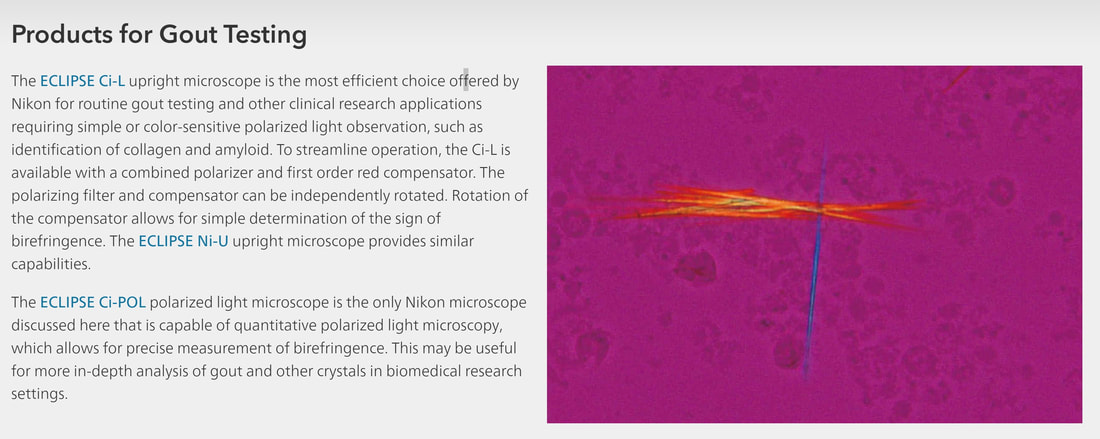
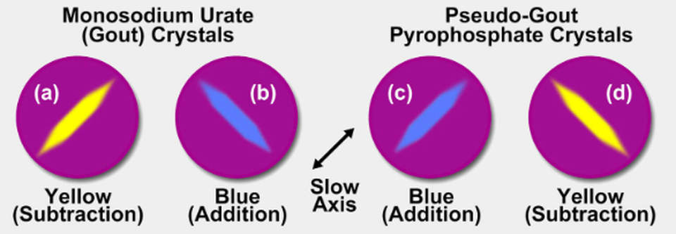
Gout crystals are generally long and needle-like while pseudogout crystals are more rhomboid. Gout crystals are negatively birefringent and their long axis is the “fast” axis (corresponding to the extraordinary wave in this case) – light polarized in a plane parallel to the fast axis will pass through the crystal faster than light perpendicular to it because it experiences a lower RI. Pseudogout crystals are positively birefringent, their long axis is the “slow” axis instead.
A polarized light microscope configured for gout testing features a linear polarizer oriented at 0 degrees (east-west) following the light source. When the long (fast) and short (slow) axes of the gout crystal are oriented at ±45 degrees, the energy of the incident light is split equally between these directions. A phase shift results as these two rays traverse the crystal due to the light taking longer to travel via the slow axis.


